Retrothyroidal intramuscular haemangioma (Feb.2019)
Case presentation
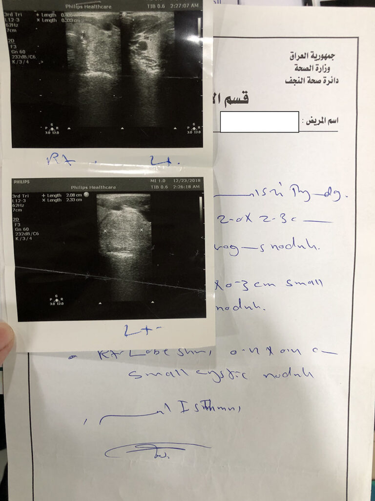
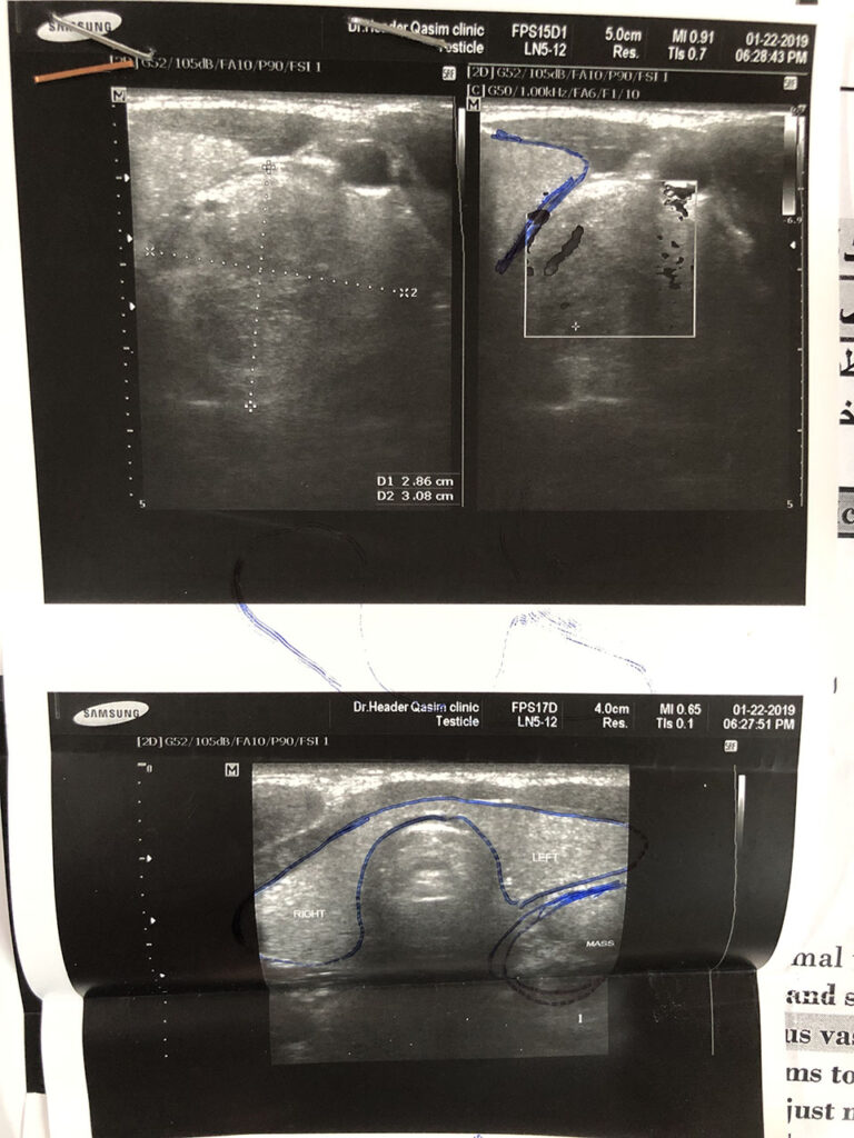
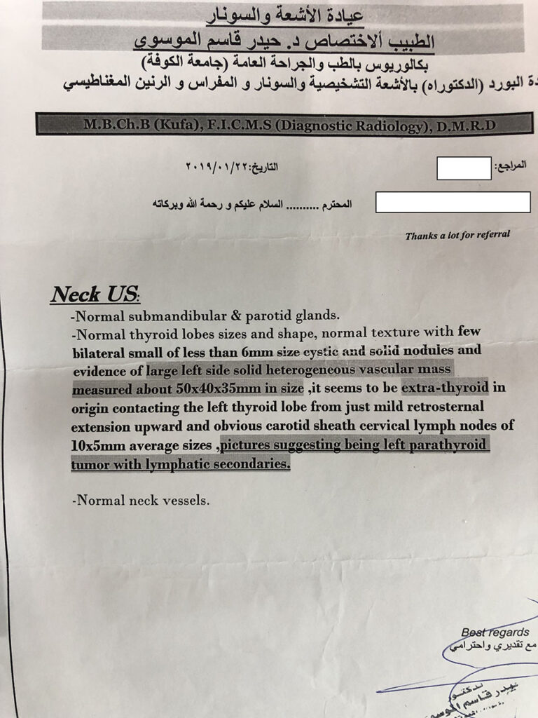
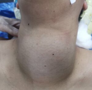
A 16 years old female presented in private clinic with 5×3 cm painless mass in Left anterior triangle of neck. On clinical examination the mass was firm fixed and didn’t move with swallowing. Ultrasound examination showed 5x3x2 cm vascular swelling in left anterior triangle posterior to the left thyroid lobe. Thyroid gland was normal.
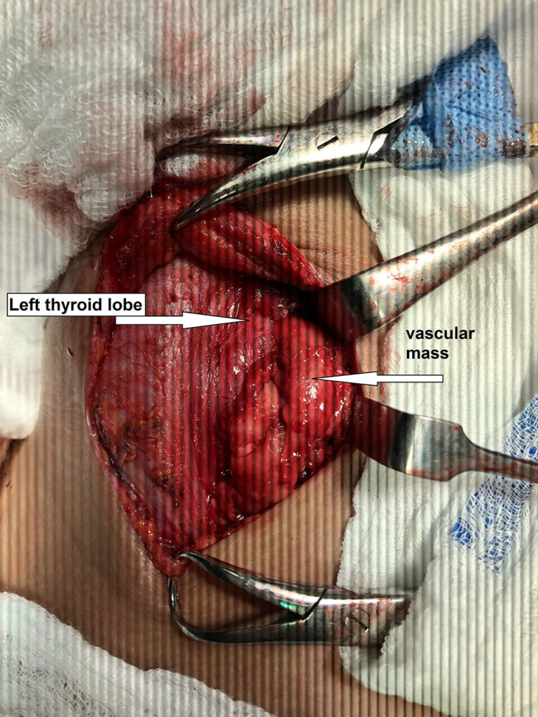
The mass was just behind thyroid lobe mimicking parathyroid mass.
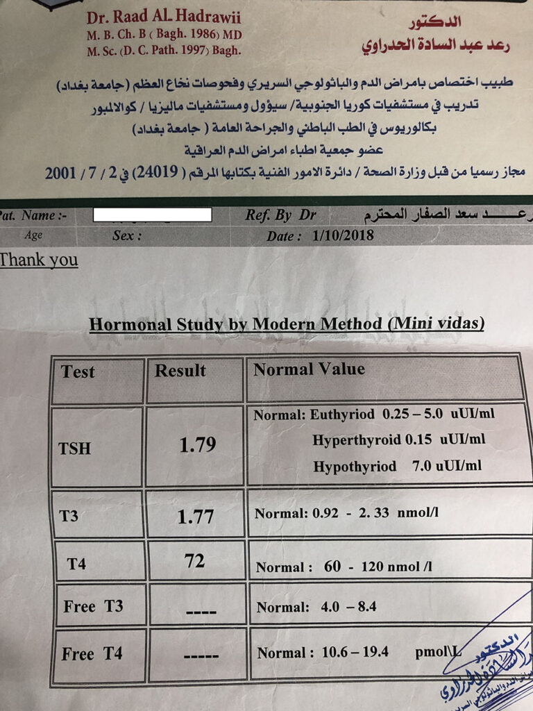
Thyroid function test was normal. Serum calcium and parathyroid hormone were within normal.
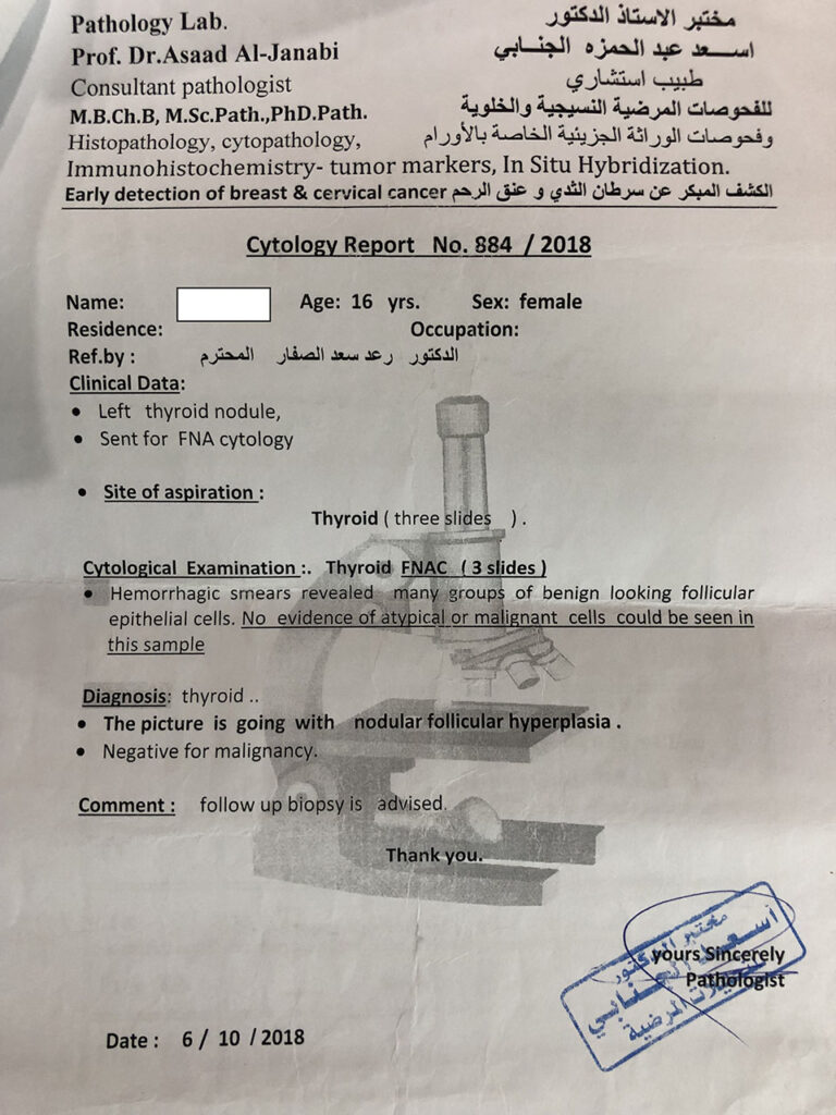
Provisional diagnosis of parathyroid adenoma was made and the patient was scheduled for operation.
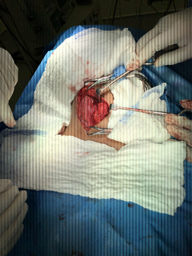
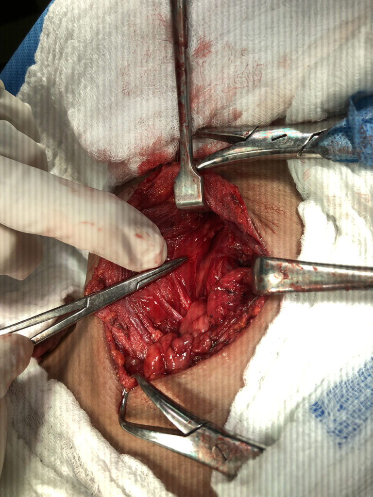
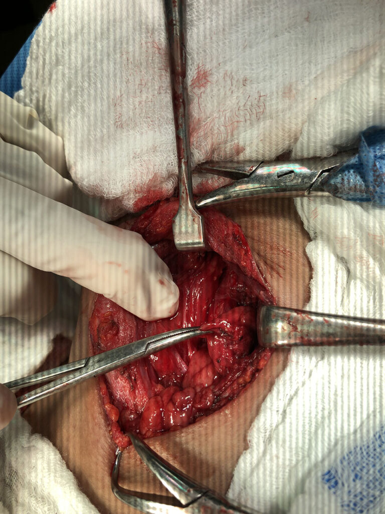
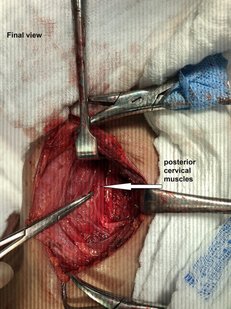
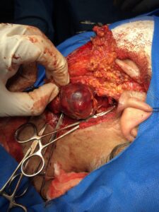
On exploration the mass was posterior to left thyroid lobe but not attached to it. It was attached to the posterior paravertebral muscle and needle aspiration revealed fresh blood.
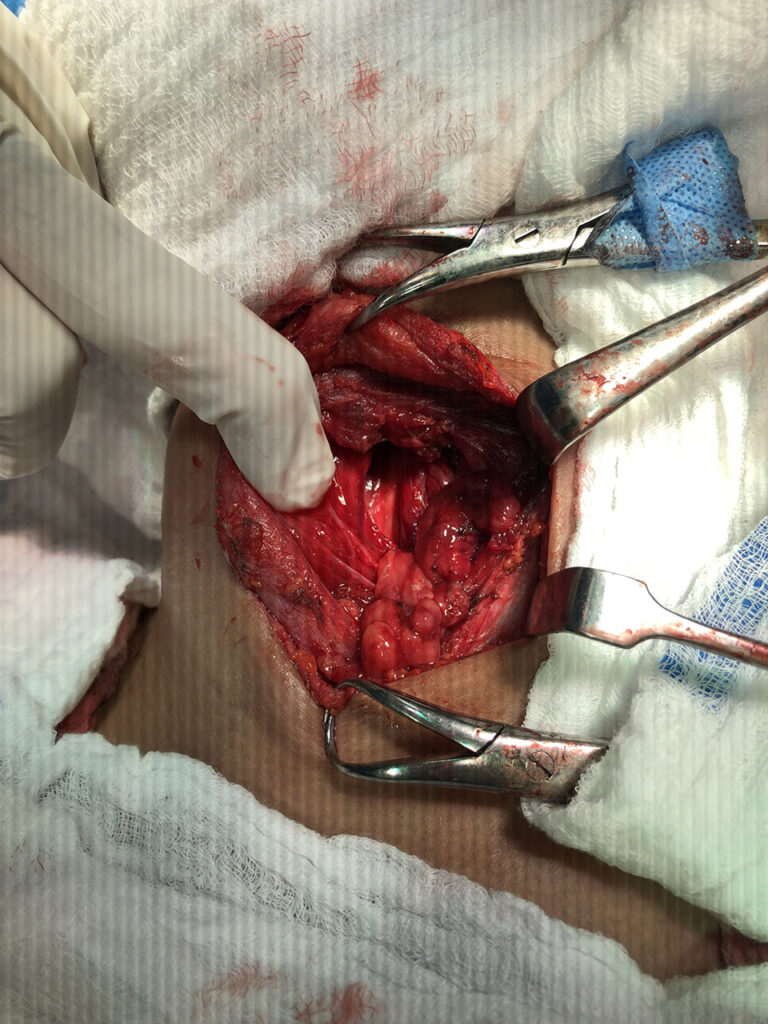
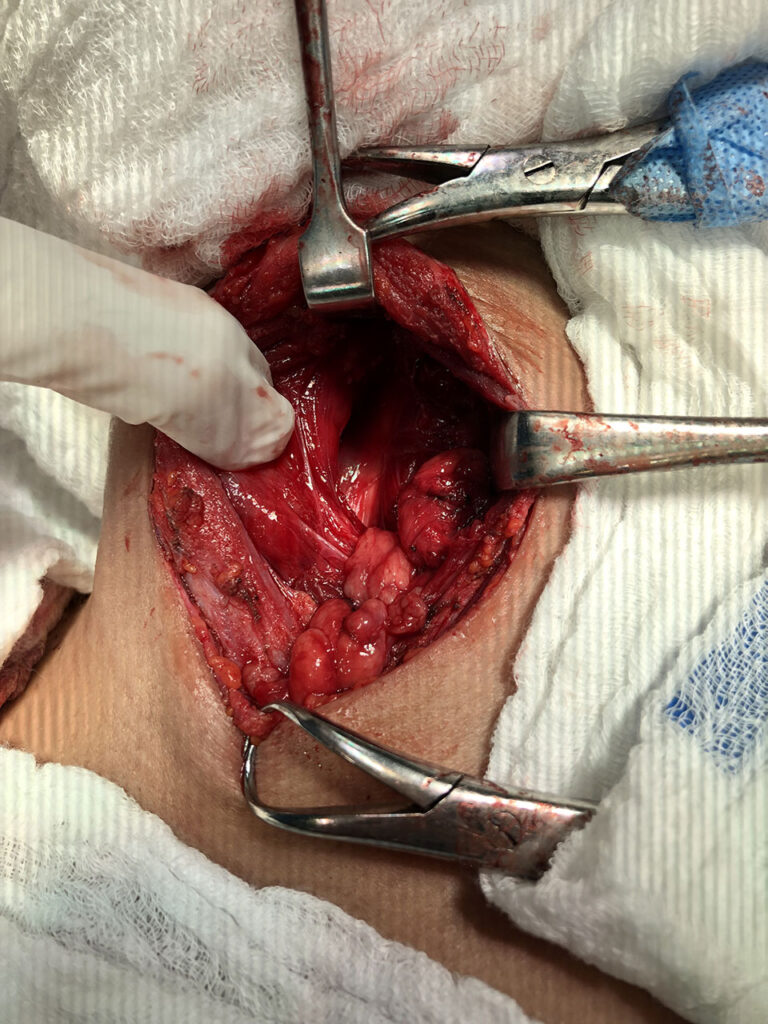
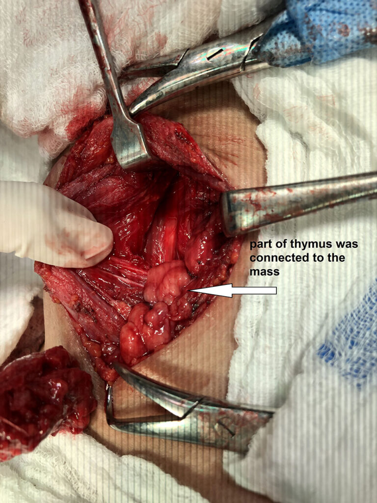
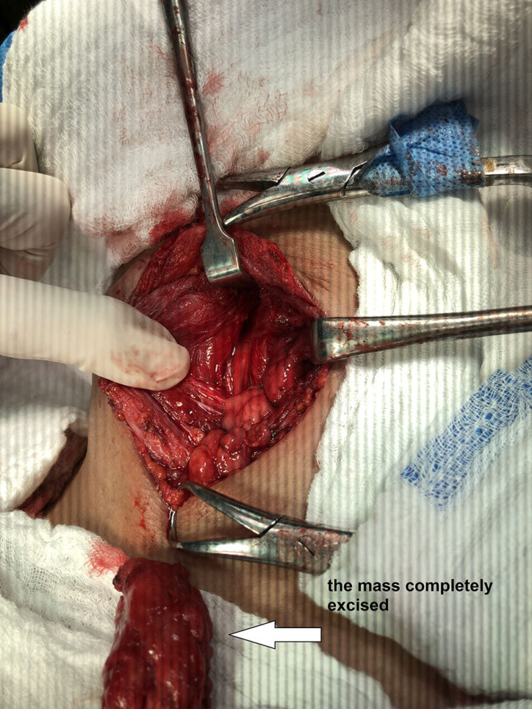
The mass was totally extracted with unusually located thymic tissue which excised with the mass.
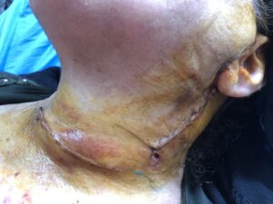
Hemostasis done and the final view was dry however tube suction drain inserted and wound closed in layers.
The postoperative course was uneventful and patient discharged well on first operative day.
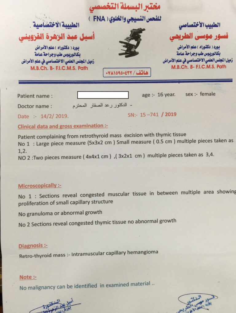
Histo pathological analysis revealed that it was an intramuscular capillary hemangioma; of rare location which was the first case to me in last 15 years.
A rare case of cavernous hemangioma of neck mimicking lipoma
Aditya Parimal Lad*, Paras Batra, Iresh Shetty, Ishant Rege, Gaurav Batra, Akriti Rajkumar Tulsian, Saurabh Mahesh Thakka
Abstract
Intramuscular hemangiomas of the head and neck are rare congenital vascular tumors and are sparsely reported. Hemangiomas account for approximately 7% of benign tumors and usually present as a mass that suddenly enlarges. Hemangiomas are mostly seen on the trunk and extremities, but can also appear on the head and neck region. A 28 year old female presented in OPD with 5?4 cm mass in Right posterior triangle of neck. CT scan showed 5x4x4 cm swelling in right posterior triangle involving sternocleidomastoid muscle. The mass was totally extracted by surgical intervention and pathological analysis revealed that it was a cavernous hemangioma.
